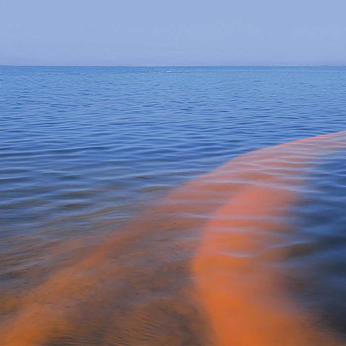
In 2008-2009, a deadly algae bloom struck the Arabian Gulf. The cluster of Cochlodinium polykrikoides was so large it threatened millions of residents, the ecology of the waterway itself and even national security.
In the years since, smaller blooms, also called red tides or harmful algal blooms, have hit the Gulf region. And warming waters seem to assure that such events will become larger and more frequent in the Middle East and around the world.
Algae aren’t inherently bad, though, says Shady Amin, an associate professor of biology at New York University Abu Dhabi who studies the phenomenon in the UAE and the Gulf of Mexico.
LISTEN TO THIS STORY HERE.
“Algae are actually for the most part good and important,” he tells KUST Review. “Without algae there would not be life on Earth. They make the oxygen we breathe. All the fish we eat is because of algae. There are good blooms that happen in the open ocean. If you go to the North Pacific, for example, very well known spring blooms enrich the food web and that’s how you get whale-watching.”
But bad blooms? The kind fed by warming waters and runoff from human activity like construction and agriculture? They can be catastrophic.
HUMAN HEALTH
Humans don’t even have to be in the water to be harmed by a red tide.
The algae can produce toxins that build up in the food chain, so humans who consume the shellfish and other fish that eat these algae face serious health risks, Amin says. “Not to mention that in areas that have a lot of wave action, a lot of these toxins can get spread into aerosols that people can inhale as they walk across a beach.”
WHEN IS A RED TIDE NOT RED?
Harmful algal blooms are often called red tides, but they can be other colors as well. Read more›››
It’s not just walking across a beach that can expose people to these risks. A study in the U.S. state of Florida found a spike in hospital visits involving respiratory distress during red tides.
And some species may not be toxic, but they can still produce irritants that affect skin and eyes. That was the case in 2018 when Abu Dhabi authorities closed local and tourist favorite Saadiyat Beach to swimmers.
ECOSYSTEM DAMAGE
Fish and other marine animals suffer as well. Get a big enough bloom, and the algae consume the oxygen in the water, creating dead zones that suffocate other sea creatures.
Animals not in those dead zones can still be injured or killed, like humans, by consuming fish or shellfish tainted by the algae’s toxins.
The results can be devastating.
The 2008-2009 event off the UAE coast, for example, wiped out thousands of tons of fish and damaged coral reefs, according to researchers who studied it.
And a 2018 red tide left Florida beaches littered with rotting fish, eels, porpoises, turtles and one nearly 8-meter whale shark. Also in Florida the same sort of respiratory problems that affect humans can afflict manatees, a threatened species, sometimes killing them. The toxins can even produce a foam that fatally strips the weatherproofing from seabirds’ feathers.
ECONOMY AND NATIONAL SECURITY
Human industries suffer as well, and not just the fisheries that rely on healthy wild and farmed stocks in the ocean.
In 1995, a C. polykrikoides bloom lasting eight weeks along the entire south coast of Korea resulted in economic losses totaling U.S.$95 million. Refineries and other coastal industries take a hit when blooms clog seawater-intake systems.
Tourism, too, suffers when locations sold as escapes to the sun and the sea become unsafe – and unpleasant – to visit. That 2018 red tide in Florida cost the state about U.S.$2.7 billion in tourism revenues, according to a study from the University of Central Florida.
But the UAE, which relies on desalination plants along its coast to supply most of its population with clean water, faces additional threats if harmful algal blooms force the plants’ closure.
This happened in 2008-2009, when the red tide struck the Arabian Gulf, forcing Gulf Cooperation Council governments to order desalination plants to shut down. The 70 plants in the UAE provide most of the country’s potable water. And this alarms Athol Yates, who keeps an eye on civil-security matters as part of his work at Khalifa University.
“If you shut down the desalination plants, normally you have a supply for a day or two,” he says. “In general, virtually the only water you’ve got is in the water-distribution network, which starts with the desalination plant and ends with your tap.
“Of course, the bulk of that water is not used for drinking purposes but (for things like) watering gardens. If you can get a message out saying, ‘Do not use the water for any other purpose, don’t wash your car,’ it will last longer. But the issue is how long will that message take to get out,” Yates says.
The country has mitigation measures in place, he adds. “The government has built a water network with redundancy, like an electricity network. So if (nuclear power plant) Barakah goes down, you still get electricity from other sources. Same with the water system.”
There is also strategic water storage, Yates says, pointing to a depleted aquifer in Liwa that has been refilled to become a below-ground reservoir. “If bad things happen to the desalination network, they can pump the water there to supply the networks.”
It’s a limited resource, however. And it’s an expensive solution. The Abu Dhabi Water Resources Master Plan estimates that desalination plants’ decreased production during the red tide cost the industry more than U.S.$100,000 (Dh368,000) a day.
GULF CONSIDERATIONS
The UAE and the Gulf have other special concerns when it comes to harmful algal blooms.
For one, Yates points out, the current around the Gulf is particularly slow, meaning a red tide might linger longer.
“These long, slow blooms can shut down the (desalination plants’) intake across a large area,” he says. “When you think about it, the (distance from) Abu Dhabi to Ras Al Khaimah isn’t that long.”
NYU Abu Dhabi’s Amin has seen how long blooms can linger. His team watched “a really nasty bloom” in Abu Dhabi’s Yas Bay for about a year. The source is uncertain, but Amin suspects runoff from construction projects or other human activity.
Amin also notes that the Arabian Gulf in general is highly oligothrophic, meaning it is nutrient-poor.
“We came to realize this very recently,” he says. “What that means is there’s not a huge amount of biomass in the water relative to areas that have a lot more nutrients. So if you have any kind of disturbance of that system, any runoff water, from, say, development or agricultural land or a monsoon, or some kind of weather event, that can really disturb the system and suddenly you get a bloom. These things are highly unpredictable.”
The Gulf also makes observing blooms difficult.
“One of the easiest things that people use is satellite imaging. It’s free. And it’s always there. Unfortunately it doesn’t work here very well because the Arabian Gulf, especially the UAE coast, is extremely shallow,” Amin says.
“Satellites measure light reflected off the Earth. And when you’re trying to approximate if there’s a bloom in a given part of the ocean, you’re measuring the wavelength that’s reflected off the water and there are algorithms that can calculate how much algae is in the water. The problem is that if you have a shallow area, you have also light reflecting off the bottom. It’s much more complicated in that case, and that’s exactly the problem we have here. So relying on satellites here is not an option.”
WATCHING THE WATER
Florida invests heavily in its red-tide defenses, Amin says.
“We work closely with the Gulf of Mexico and the State of Florida. They have a program funded by the state with hundreds if not thousands of state employees that go out almost every day, and they collect water. If it matches a certain threshold that they know a bloom is happening, they alert the public and start taking action.”
The UAE does not yet have these kinds of resources, Amin says, “but we’re making progress.” “That’s what we aim to do.”
Oman, meanwhile, in early 2024 launched a predictive model to help warn of incoming red tides.
An early warning system is indispensable for countries in the region, especially as climate change could make red tides more frequent and deadly, says Jauad el Kharraz, head of research at Muscat-based Middle East Desalination Research Center.
He stresses that further research is vital to evaluate the relationships between red tides, climate change, ocean acidification and human health.
Amin, meanwhile, suggests that we should be looking closer at ourselves. “It’s only because of us overdeveloping our coasts and dumping things in the sea water that leads to these harmful red tides,” he says.
A BETTER FILTER
By: Jade Sterling
High levels of nutrients sounds like a benefit to an ecosystem, but when an environment receives too many nutrients, otherwise known as eutrophication, algal blooms and hypoxic waters can kill fish and seagrass.
“The high accumulation of nutrients, including nitrogen and phosphorus, discharged into surface water, rivers and reservoirs can accelerate eutrophication and cause great damage to the aquatic ecosystem,” says Shadi Hasan, director of the Center for Membranes and Advanced Water Technology at Khalifa University.“We need to control the levels of nutrients and develop innovative technologies to treat water and remove excess nutrients.”
Novel membrane technology, however, may be the solution. Hasan’s KU research team has developed a composite polylactic acid (PLA) and nanomaterial membrane to remove nutrients from wastewater.
Treatment technologies already exist. However, chemical methods can introduce undesirable byproducts; and biological treatments take much longer and are inefficient in the use of nitrogen. Additionally, no available method offers complete water purification.
The membrane works via adsorption. The research team used a functionalized positively charged multi-walled carbon nanotube/graphene oxide hybrid nanomaterial to remove nitrogen (as ammonia) and phosphorus from wastewater while enhancing water permeability. The nutrients are filtered out by collecting in the pores of the nanotubes at the surface of the membrane.
But such a membrane needs to offer water permeability. As more nutrients adsorb and collect, the amount of water passing through decreases. The research team’s membrane, however, offers high water flux even when filtering the nutrients.
The carbon nanotubes increase membrane tensile strength significantly, while the graphene oxide enhances thermal stability and tensile strength and provides antibacterial properties. This supports water flux and provides hydrophilicity to the end product.
While the effects of graphene oxide and carbon nanotubes in water purification are well-documented, studies are limited when it comes to combining the two as a nanohybrid.
“After a comprehensive review of the literature, our research group is the first to report the fabrication of such composite PLA membranes for the removal of nutrients from synthetic and real wastewater,” says Hasan, who adds that the team is investigating ways to scale up the membranes for larger applications.
More like this: High seas