

Khalifa University professor teams with industry pros
and Nobel laureate to improve strategy›››
Health
Have you ever been playing around with Google Earth just to see if your childhood home is still standing?
Researchers from University College London and the European Synchrotron Radiation Facility created something similar, but for human organs — not to make sure they’re still there, but to explore them on an unprecedented scale. And anyone can use it.
Inspired by structural lung damage resulting from SARS-CoV-2 and the lack of imaging to help researchers understand how this happens, the team created a database of 3D organ imaging called the Human Organ Atlas. Users can zoom in to view the organ at a near cellular level, no cuts required.
This online library is free to use and contains 3D scans of real human organs. It allows an immersive learning experience for students and researchers alike.
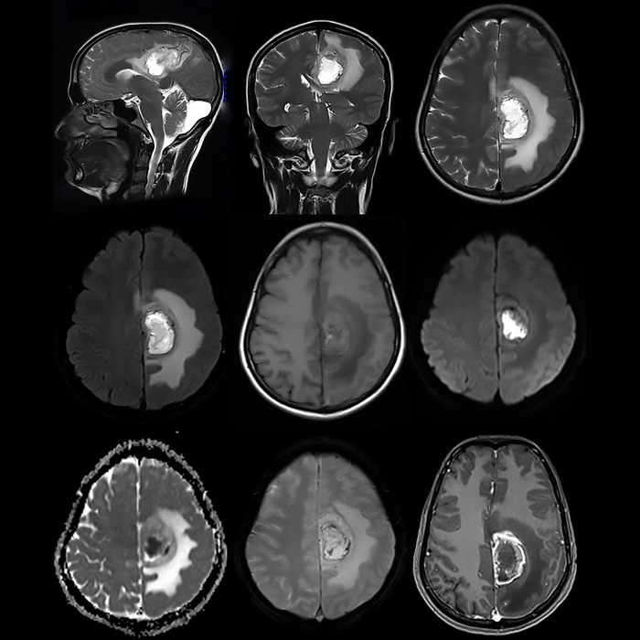 CAPTION: Glioblastoma MRI Image IMAGE: Shutterstock
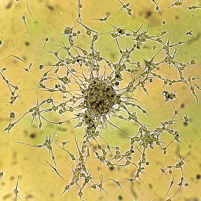 CAPTION: Glioblastoma cell culture IMAGE: Shutterstock
The tool can offer insight into disease pathology to help to build better treatments and medicines and a completely immersive anatomy lesson. It is also expected to contribute greatly to the development of AI medical systems.
Three-D organ scanning has been around for some time but many people were frustrated to zoom in to find blurry images that make fine details difficult to see. Additionally, tissue samples from organs in detail could be viewed under a microscope, but there was no way to view the organs and the tissue cells synchronously. Now both can be studied as a complete system.
The atlas was built using scanned organs from autopsies and a synchrotron (particle accelerator).
A synchrotron, roughly the size of a football stadium, “accelerates electrons very, very fast. And as these electrons are bent with magnets, they give off X-rays. And it’s these X-rays that we’re using for imaging,” Claire Walsh, lead author on the paper and director of the Human Organ Atlas Hub, tells Science Friday.
Currently we work on isolated organs, but in the future, we expect to develop the technique to be able to image complete human bodies with a resolution 10 to 20 times higher than what is possible today. Such data could transform how anatomy is studied and understood.”
– Paul Tafforeau, Beamline scientist, European Synchrotron Radiation Facility
The scanning technique called hierarchical phase-contrast tomography (HiP-CT), was developed in 2021 by Walsh and her team. It scanned at about 20 microns per voxel — that’s roughly thinner than a human hair. The scans are 100 billion times brighter than conventional hospital CT scanners.
There are currently 62 organs from 12 organ types in the atlas: the brain, heart, lung, kidney, liver, colon, spleen, placenta, uterus, prostate, testis and eye. These organs offer insight into conditions such as hypertension, cancer, damage from COVID-19 and rarer disorders like Dandy-Walker Syndrome.
The world-wide usable atlas offers downloadable datasets (in multiple resolutions), tutorials and software tools for analysis, ongoing data additions and interactive browser-based visualization.
This level of biomedical imaging has been a goal for decades, and it was declared officially a functional whole-organ imaging technique in 2021. The online Human Organ Atlas was launched for all users, whether you’re a researcher, student or just an interested individual, in March 2026.
More like this: Teamwork makes the dreamwork
Get the latest articles, news and other updates from Khalifa University Science and Tech Review magazine